 |
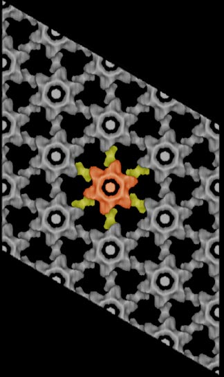
A three-dimensional reconstruction of the murine
leuekemia virus capsid protein was derived by electron microscopy and image
aalysis of two-dimensional crystals. An individual hexamer has been colored
red and yellow to highlight the two capsid domains within the lattice. The
yellow domains are linkers between adjacent capsid hexamers. Recent analyses
of other retroviral capsid proteins (HIV-1 and Rous sarcoma virus) have
demonstrated a similar arrangement of capsid molecules within almost identical
hexameric lattices, suggesting a general model for retroviral assembly.
|